
Amsler Grid Test
Table Of Contents
These are some of the questions we were asking ourselves, so we decided to make this article to clarify these and other doubts related to metamorphopsia and the Amsler Grid for its early detection.
- What is the Amsler Grid Test?
- What does the Amsler Grid test evaluate?
- How is the Amsler Grid test used?
- How to interpret the results of the Amsler Grid test?
- Who was Marc Amsler?
- What is the interactive Amsler Grid?
- What causes metamorphopsia, and how is it related to the Amsler Grid?
- Do we have distorted vision, or do we see ripples?
In addition, at the end of the article, we have attached a PDF with the Amsler Grid Test, a downloadable application for cell phones, and a link that will take you to an online metamorphopsia test.
What is the Amsler Grid?
The Amsler grid tests the central areas of the eye’s visual field. It is a method of self-assessment and rapid detection of defects in the central or macular field of vision, such as age-related macular degeneration or other retinal-related degenerative processes.
The Amsler test is a test that only evaluates the central retina, so it is still required to perform other ophthalmologic examinations with dilated fundus to assess the peripheral retina as well.
The Amsler test was created by the Swiss ophthalmologist Marc Amsler in 1945 and is performed when there is any suspicion of a problem in the macula, seeing undulations, twisted lines, or distorted vision.
The macula is a small part of the central retina responsible for your vision’s ‘sharpness’ and ‘resolution.‘ It takes care of your central vision or straight-ahead vision, thanks to a large number of cone photoreceptors.
The Amsler grid test aims to detect pathologies affecting the center of the retina, the macula.
Who was Marc Amsler?
Marc Amsler (February 5, 1981, Vevey, Zurich – May 3, 1698) was a professor of ophthalmology at the University of Zurich Ophthalmology Clinic from 1944 to 1961, after being chief ophthalmologist in Lausanne since 1935.
Dr. Amsler is known for his ‘Amsler Grid,’ which improved the work initiated by ophthalmologist Edmond Landolt (known for Landolt C Test)
Professor Marc Amsler also performed the first corneal transplantation in 1951 at the Zurich Ophthalmology Clinic. He has also published several papers on keratoconus and uveitis. Among others:
- Earliest symptoms of diseases of the macula. The British Journal of Ophthalmology, London, 1953, 37: 521-537.
- Fuchs’ heterochromia and vascular fragility (with Florian Verrey). Ophthalmologica, Basel, 1946, 111: 177.
- Le kératocône fruste au Javal. Ophthalmologica, Basel, 1938, 96: 77-83.
Lehrbuch der Augenheilkunde. Basel, Karger, 1948. 858 pages. M. Amsler, A. Bruckner, Adolphe Franceschetti, Hans Goldmann u. Enrico Bernardo Streiff, editors: New edition 1954, 927 pages.3rd ed, Basel, Freiburg im Breisgau, New York : Karger, 1961. 1011 pages. - L’Humeur Aqueuse et ses Fonctions (with Florian Verrey and Alfred Huber). Paris, Masson, 1955.
- Mydriase et myose directes et instantanées par les médiateurs chimiques. with Florian Verrey. Annales d’Oculiste, Paris, December 1949, 182 (12): 936.
- Quantitative and qualitative vision. Transactions of the Ophthalmological Society of the United Kingdom, London, 1949, 69: 397-410.
How To Perform An Amsler Grid Test?
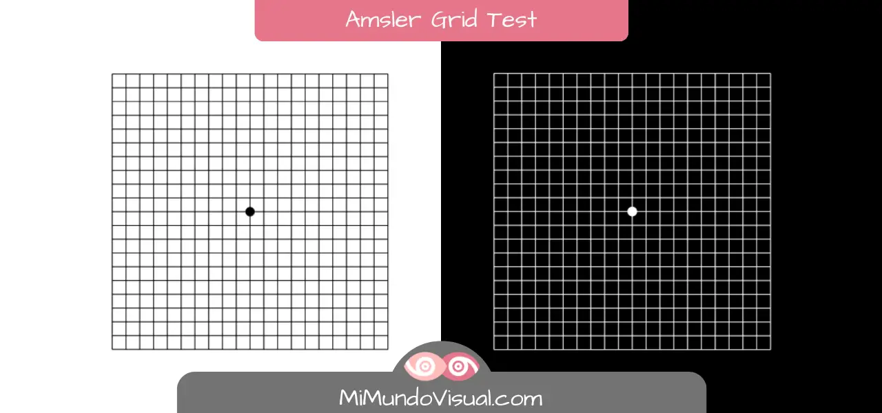
The Amsler grid consists of a 10 cm x 10 cm grid presented on a black background (it is also possible to perform it on a school blackboard). With a dot in the center, each square of the Amsler grid measures 5 mm and consists of parallel, vertical, and horizontal white lines. Each grid square has an angle of 1º degree at a 28 – 30 cm distance, and the whole grid is 20º high by 20º wide.
The person performing the test with the Amsler grid should cover one eye with the palm of the hand or with an eyepatch since it is a monocular test (i.e., with one eye at a time). And it will be placed at a distance of 28 – 30 cm.
If the person notices dark spots, holes in the grid, waves or curvatures, or identifies central shadows, an ophthalmological visual examination should be performed as soon as possible.
Even if you have performed the Amsler grid test without having any of these visual perceptions, it is not entirely ruled out that you do not suffer from any defect in the central or peripheral field of vision. Therefore, it is important to remember to have regular vision examinations.
The 7 Amsler Grid Charts
There are 7 different Amsler Grid charts. These consist of 7 sheets with slight variations of the original Amsler Grid:
- The most commonly used Amsler Grid:
It is a white grid with a black background, a white center dot, and a total of 20 5 mm squares. - White grid with two diagonal lines in addition to the grid:
It is used to detect central scotomas. - Red grid with black background:
Used for color scotomas. Red stimulates long wavelength cones. - Grid with random white dots:
Instead of lines with a black background and a central white dot, it is used to detect scotomas. - A grid with 21 parallel white lines: spaced every 5 mm apart with a black background and a central white dot for fixation. It is used to detect metamorphopsia and reading difficulties.
- Grid with black parallel lines on white background:
With less distance between them in the grid, used to detect fine metamorphopsia near fixation. - Grid with smaller squares in the central area: each with a field of 0.5°, is similar to the grid of the first slide, with the difference of having smaller squares. It detects subtle changes in the early stages of pathologies with alteration of the macula.
Amsler Interactive Grating (Amsler Digital Grid)
The digital Amsler grid is an electronic version of the Amsler grid that allows the progress of the Amsler grid to be monitored quantitatively as opposed to the original Amsler grid.
The person performing the Amsler test with the interactive Amsler grid must straighten the crooked lines with the help of the computer mouse or tactilely.
In this way, it is possible to have an interactive measurement of metamorphopsia. This quantitative method allows for calculating a 3D image of the distorted perception of the person performing the Amsler digital grid test.
Amsler Grid Interpretation
The interpretation of the results of the Amsler grid test is simple. If, during the Amsler test, the person can see the edges of the Amsler grid and the vertical and horizontal lines, the results are satisfactory.
Any different result will mean an alteration in the macula which refers to an anomaly in vision.
The Amsler grid test allows for detecting ocular pathologies related to changes in the central retina and helps to detect them early.
Causes of Wavy Vision
Seeing ripples or shaky vision is due to alterations in the macula that can be seen by performing the Amsler grid test.
Therefore, with the Amsler grid test, it is possible to detect eye diseases associated with the macula.
What is Metamorphopsia?
The definition of metamorphopsia in ophthalmology refers to a vision with altered or distorted images.
Ocular metamorphopsia is not a disease but a symptom. It is a symptom associated with some pathology affecting the macula located in the center of our retina.
Distortion of vision or metamorphopsia refers to an alteration in the perception of people or objects in both shape (dysmorphopsia) and size (dysmegalopsia), in addition to seeing undulations and crooked lines when they are actually straight.
Although it is a binocular disorder that affects both eyes, it will first affect one. It may take a long time before we are aware that we have a distorted vision since the brain integrates the two images it receives in the brain. The distortion is not perceived when it is small.
Amsler Grid and Metamorphopsia
The diagnosis of ocular metamorphopsia is made utilizing the Amsler grid test.
Suppose when observing the Amsler grid, the person sees crooked lines or undulations in any part of the Amsler grid. In that case, they will know that they are suffering from a distortion in the visual field at that same point.
Causes Of Metamorphopsia
The causes of ocular metamorphopsia are:
- Diseases of the central part of the retina, the macula. Some of the ocular pathologies that can cause metamorphopsia are age-related macular degeneration (AMD), macular degeneration due to myopia magna, histoplasmosis syndrome, central serous chorioretinopathy, diabetic macular edema (DME), deposits in the macular area, obstruction of the retinal blood vessels, macular dystrophies, tumors in the macula, uveitis, epiretinal membrane, etc.
- Neurological diseases such as epilepsy or migraine can also cause metamorphopsia or distorted vision.
As well as any medication containing any psychoactive substance.
Early Detection of Age-Related Macular Degeneration AMD Using the Amsler Grid
Age-related Macular Degeneration AMD is a degenerative pathology affecting the macula. It can be detected using the Amsler grid test.
The person suffering from AMD disease, when performing the test, will not see straight lines but deformed, cloudy, wavy, or even incomplete lines. It is possible to see the central point as a spot in the middle of the grid.
The AMD disease involves progressive vision loss, is irreversible, and can only be slowed down. Therefore, it is essential to go to the ophthalmologist when you notice any vision loss or when you see distorted elements around you.
For more information on Age-Related Macular Degeneration AMD, you can read the Protocol for diagnosis, follow-up, and general recommendations in early and intermediate age-related macular degeneration (AMD): consensus of a panel of experts.
Treatment For Metamorphopsia
Treatment for metamorphopsia will depend on the pathology causing the distorted vision.
Metamorphopsia is not a disease but a symptom of a disease of the retina’s center. Therefore, the cure for metamorphopsia will depend on the disease causing it.
The Amsler test is used for the early detection of retinal pathologies. Early detection is of vital importance to prevent or treat total loss of central vision.
Therefore, it is very important to have regular ophthalmologic check-ups to detect any changes in the macula or center of the retina.
Patients suffering from metamorphopsia are advised to perform the Amsler grid test at home once a week to keep track of the evolution and to be able to detect any change in time.
For more information on the first symptoms of macular pathologies, you can read this interesting article by Marc Amsler himself.
Amsler Grid Test PDF
Instructions for Performing the Amsler Grid Test
- The room in which we are must be sufficiently illuminated.
- We place ourselves at 28 – 30 cm from the Amsler Grid Test.
- If we use glasses or contact lenses, we should perform the test by keeping them on.
- We perform the test by covering one eye first and then the other since it is a monocular test (one eye at a time).
- We stare at the point in the central area of the Amsler grid, having the entire grid in our field of vision.
We answer the following questions:
- Are the lines crossing the grid parallel and straight from the beginning to the end, especially in the center of the grid?
- Is each of the squares forming the grid equal and regular?
- If we see all or some lines distorted, bent, wavy, blurred, or disappear at some point, we will go to an ophthalmologist as soon as possible.
The Amsler Grid App
If you prefer, you can download this app to perform the Amsler grid test on your smartphone:
Amsler’s Grid App.